Microscopy is one of the most essential tools in biology, medicine, and material sciences. By allowing scientists to see structures invisible to the naked eye, microscopes unlock the microscopic world and drive discoveries in research, diagnostics, and technology. This guide provides a comprehensive overview of microscopy, helping you understand its types, how to choose the right microscope, and its diverse applications.
What is Microscopy?
Microscopy is the technique of using a microscope to observe objects that are too small to be seen by the naked eye. Microscopes magnify samples, providing a closer look at cells, tissues, microorganisms, and nanostructures. Modern microscopy has evolved from simple optical devices to advanced digital and electron-based systems, enabling unprecedented detail and analysis.
How to Choose the Right Microscope
Selecting the correct microscope depends on your research goals, sample type, and budget. Here’s a step-by-step guide:
1. Identify Your Sample
- Biological samples: Cells, tissues, bacteria, and viruses.
- Material samples: Metals, crystals, polymers, and nanomaterials.
- Live samples: Require gentle handling to maintain viability.
2. Determine the Required Resolution
- Resolution is the microscope's ability to distinguish two close points as separate.
- For cellular imaging, a resolution of ~200 nm is sufficient.
- For subcellular or molecular structures, higher resolution techniques like electron microscopy are needed.
3. Consider the Magnification
- Low magnification (4x–40x): Ideal for overview and tissue examination.
- Medium magnification (100x–400x): Good for cell-level observation.
- High magnification (1000x+): Required for subcellular organelles and microorganisms.
4. Sample Preparation Requirements
- Some microscopes need thin slicing or staining.
- Live cell imaging requires minimal disturbance.
5. Budget and Maintenance
- Optical microscopes are cost-effective and user-friendly.
- Confocal and electron microscopes are expensive and require trained personnel.
Types of Microscopes and Their Differences
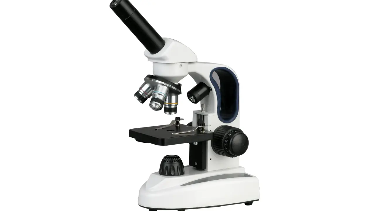
1. Optical (Light) Microscopes
Uses visible light and lenses to magnify samples.
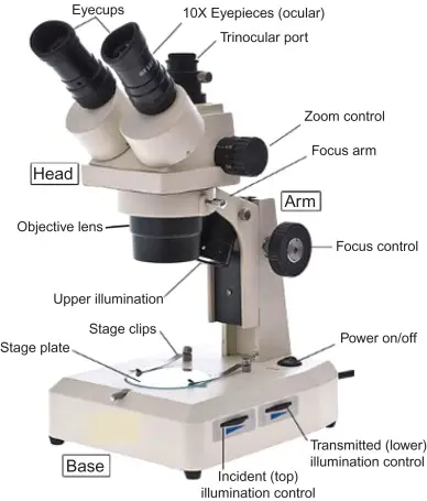
Subtypes:
- Compound Microscope: Best for thin biological samples; magnification 40x–1000x.Read more
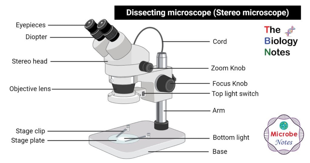
- Stereo Microscope (Dissecting Microscope): Provides 3D view; ideal for larger specimens.Read more
- Fluorescence Microscope: Uses fluorescent dyes; excellent for tracking molecules in cells.

Pros: Easy to use, affordable, versatile.
Cons: Limited resolution (~200 nm).
2. Confocal Microscopes
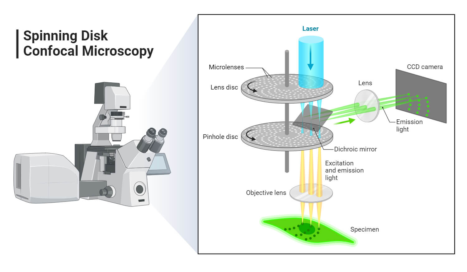
Uses laser light to scan samples and create 3D images.
Applications: Live cell imaging, tissue reconstruction, molecular studies.
Pros: High-resolution, 3D imaging.
Cons: Expensive, requires training.
3. Electron Microscopes
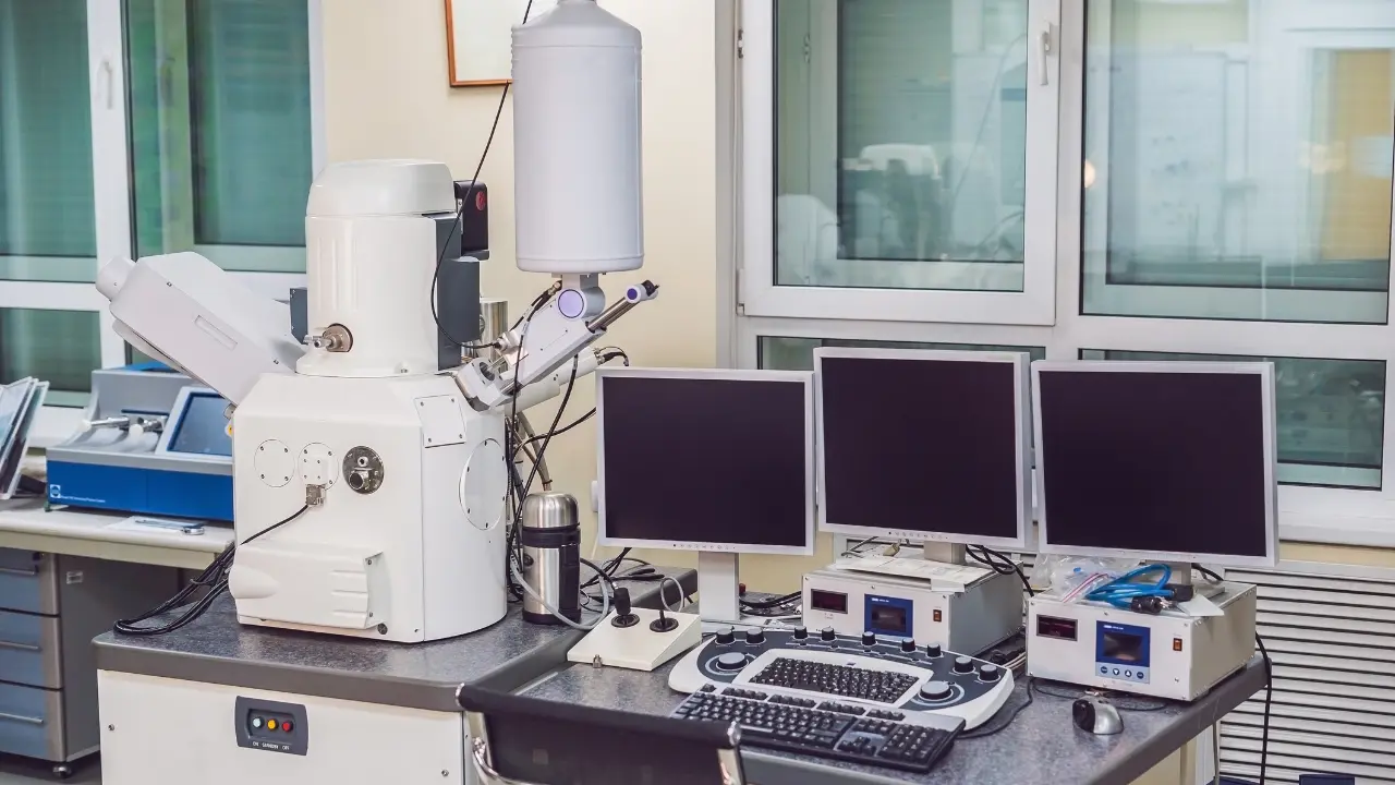
Use electron beams instead of light, achieving nanometer-scale resolution.
Subtypes:
- Transmission Electron Microscope (TEM): Ultra-high resolution for internal structures.
- Scanning Electron Microscope (SEM): 3D images of surfaces.
Pros: Unmatched resolution.
Cons: Costly, sample preparation is complex, cannot view live specimens.
4. Digital Microscopes
- Connected to computers or tablets.
- Can capture and store high-resolution images.
- Ideal for teaching, documentation, and remote analysis.
5. Other Advanced Microscopy Techniques
- Phase-Contrast Microscopy: Visualizes transparent samples without staining.
- Atomic Force Microscopy (AFM): Measures surface topography at atomic resolution.
Microscopy in Diagnostics
Microscopy is critical in medical and clinical diagnostics.
Examples include:
Hematology: Blood smear examination for anemia, leukemia, and infections.
Microbiology: Identifying bacteria, fungi, and parasites.

Histopathology: Tissue biopsy analysis to detect cancer or inflammation.
Virology: Studying virus morphology with electron microscopy.

Cytology: Pap smears and other cell-based diagnostics.
Microscopy enables rapid detection, early diagnosis, and monitoring of diseases, improving patient care outcomes.
Applications of Microscopy
Read more
Microscopes are not limited to diagnostics; they are essential across research, education, and industry:
1. Biological and Medical Research
- Studying cell biology, molecular pathways, and tissue architecture.
- Observing cellular responses to drugs or treatments.
2. Materials Science
- Analyzing metals, polymers, and nanomaterials.
- Surface characterization and quality control.
3. Pharmaceutical Industry
- Drug development and testing.
- Crystal structure analysis for drug formulation.
4. Environmental Science
- Detecting microorganisms in water, soil, and air.
- Studying ecological interactions at the microscopic level.
5. Education
- Teaching students about cells, tissues, and microorganisms.
- Hands-on experience with modern imaging techniques.
Tips for Effective Microscopy
- Proper sample preparation is crucial for clear imaging.
- Use the right objective lens for your desired resolution and field of view.
- Adjust illumination and contrast to enhance visibility.
- Regularly maintain and calibrate microscopes to ensure accurate results.
Conclusion
Microscopy is a cornerstone of modern science and medicine. Choosing the right microscope involves considering your sample, resolution needs, and budget. From basic optical microscopes to advanced electron systems, each type has unique advantages and applications. By mastering microscopy techniques, researchers and clinicians can explore the invisible world, advance scientific knowledge, and improve healthcare outcomes.